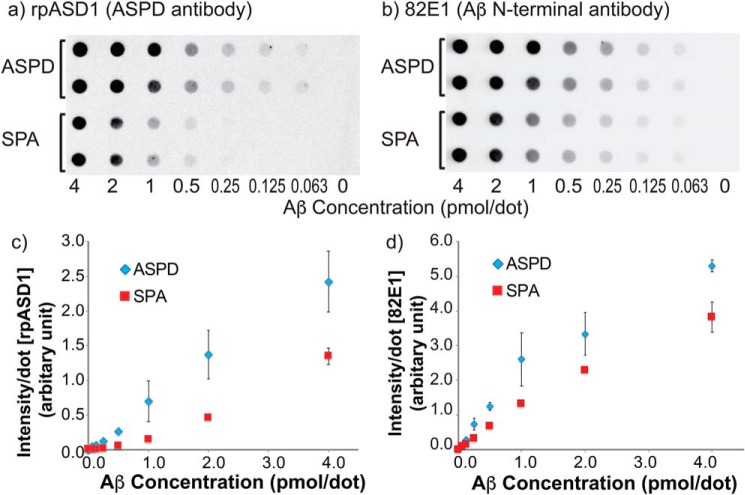
Figure 5.
a and b, dot-blot assays for different concentrations of SPA and ASPD using rpASD1 anti-ASPD antibody (a) and 82E1 antibody (b), the latter of which targets Aβ(1–16). Serially diluted samples were loaded on the dots in duplicate (a and b), and the average intensities of the dots were plotted in c and d. The rpASD1 assays (a and c) were used for the detection of ASPD species, whereas the 82E1 assays (b and d) were performed as a control for the quantification of Aβ species. Error bars, S.D.

