Figure 2.
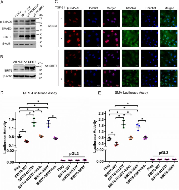
A, Western blot analysis of cardiac fibroblasts transfected with FLAG-control, FLAG-SIRT6-WT, FLAG-SIRT6-H133Y, or FLAG-SIRT6-S56Y-SIRT6. n = 3. B, Western blot analysis to confirm overexpression of SIRT6 in cardiac fibroblasts, upon infection with Ad-Null or Ad-SIRT6 for 24 h. C, representative confocal images of cardiac fibroblasts infected with either Ad-Null or Ad-SIRT6, with or without TGF-β1 treatment (10 ng/ml) for 24 h, for the indicated proteins. Scale bar, 20 μm. D and E, luciferase activity assay for TGF-β1/activin-response elements (TARE-Luciferase (D)) and α-SMA-reporter luciferase (E). Luciferase plasmid was transfected in cardiac fibroblasts, along with FLAG-control, FLAG-SIRT6-WT, FLAG-SIRT6-H133Y, or FLAG-SIRT6-S56Y using Lipofectamine 2000 for 48 h. Luciferase activity was measured with or without the treatment of SB-505124 (2.5 μg/ml) for 24 h. pGL3 basic vector was used as a negative control. n = 3. Data are presented as mean ± S.D. (error bars). *, p < 0.05. One-way ANOVA test was used to calculate p values.

