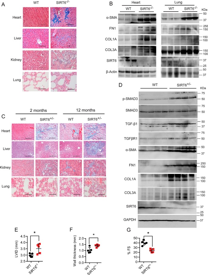
Figure 6.
A and C, histology of heart, liver, kidney, and lung tissue samples from 25-day-old WT and SIRT6-KO littermate mice (A) and 2-month-and 12-month-old WT and SIRT6+/− mice (C) showing tissue fibrosis. Scale bar, 50 μm. n = 3. B, Western blot analysis of heart and lung lysates from 25-day-old WT and SIRT6-KO mouse littermates for the indicated proteins. n = 3. D, Western blot analysis of heart lysates from 12-month-old WT and SIRT6+/− mouse littermates for the indicated proteins. n = 4 mice/group. E–G, scatterplot showing the left-ventricular internal diameter (LVID), left-ventricular posterior wall thickness, and cardiac contractile functions, as measured by fractional shortening of 12-month-old WT and SIRT6+/− mice, n = 5; data are presented as mean ± S.D. (error bars). *, p < 0.05. Student's t test was used to calculate the p values.

