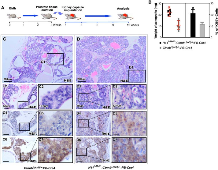
Figure 4.
Met enhances β-catenin–mediated tumor progression in xenograft model for prostate cancer. A, schematic representation of experimental design. B, graphical representation of weights of xenografts derived from H11L-Met/+:Ctnnb1(Ex3)L/+:PB-Cre4 or Ctnnb1(Ex3)L/+:PB-Cre4 mouse prostate tissues (left) or Ki67 expression (right) in prostate tissues of the indicated genotypes. Shown are representative images of H&E-stained prostate tumors from Ctnnb1(Ex3)L/+:PB-Cre4 (C) or H11L-Met/+:Ctnnb1(Ex3)L/+:PB-Cre4 (D) mice. C1–D2, high-magnification images from the indicated mice. Immunohistochemical analysis of Met (panels C4 and C5 and panels D4 and D5) and β-catenin (panels C6 and C7 and panels D6 and D7) in prostate tissues from the indicated mice. Scale bar, 50 μm (12.5 μm for high-magnification images).

