Figure 6.
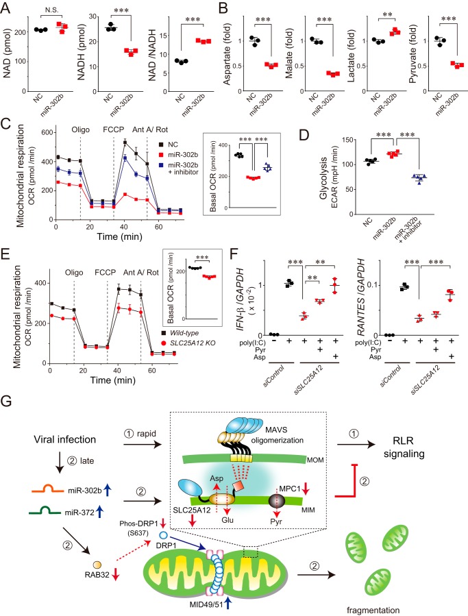
miR-302b targets SLC25A12 to disrupt mitochondrial metabolism linked to the antiviral responses. A, NAD and NADH levels (pmol/106 cells) in HEK293 cells transfected with miR-302b mimic were analyzed 72 h later by LC-MS/MS. The right graph shows the NAD/NADH ratio. N.S., not significant. Data shown represent mean values ± S.D. (n = 3). ***, p < 0.001. B, mitochondria-related metabolites in the miR-302b mimic-transfected HEK293 cells analyzed 72 h later by GC/MS. The aspartate, malate, and pyruvate levels were each significantly decreased, whereas the lactate level was increased. Data shown represent mean values ± S.D. (n = 3). **, p < 0.01 and ***, p < 0.001. C, OCR of HEK293 cells transfected with the miR-302b mimic (± inhibitor) measured using a Seahorse XFe96 Analyzer. Inset: basal OCR of each transfected cell. The injection order of oligomycin (Oligo), FCCP, and antimycin A/rotenone (Ant A/Rot) is indicated. Data shown represent mean values ± S.D. (n = 5). ***, p < 0.001. D, extracellular acidification rate of HEK293 cells transfected with the miR-302b mimic (± inhibitor) was measured using the Seahorse XFe96 Analyzer. Data shown represent mean values ± S.D. (n = 5). ***, p < 0.001. E, similar to C, except that the representative respiration of the SLC25A12 knockout and its WT HAP-1 cells was measured. Data shown represent mean values ± S.D. (n = 5). ***, p < 0.001. F, siRNA-treated HEK293 cells (siSLC25A12) were transfected with 2 μg of poly(I-C) for 24 h, and the total RNA from the cells was analyzed by qPCR for the expression of IFN-β, RANTES, and GAPDH (internal control). At the time of the poly(I-C) transfection, either pyruvate (Pyr, 2.5 mm) or aspartate (Asp, 10 mm) was added to the medium of the siSLC25A12-treated cells. Data shown represent mean values ± S.D. (n = 3). **, p < 0.01 and ***, p < 0.001. G, model of miRNAs that control RLR-mediated antiviral signaling. Cellular innate immune responses to RNA virus infection result in the rapid activation of signaling molecules, including MAVS oligomerization (inset) and the formation of signalosomes on the MOM, ultimately facilitating the RLR-signaling pathway (➀). In this process, we assume that SLC25A12, as a part of the PHB interactome (inset, highlighted with blue background) (28), interacts with MAVS in mitochondria that have a critical role in MAVS oligomerization. At longer times post-infection (∼24 h), miR-302b (and also miR-372) is up-regulated in the cells, which promotes mitochondrial fission through the DRP1-dependent pathway and also impairs mitochondrial metabolism (➁). We propose that the miRNAs undergo sequential negative regulations following viral infection, a process that is critical for terminating excess RLR signaling.

