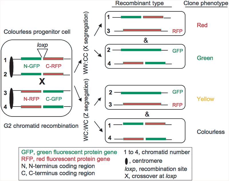
Fig. 2.
The mouse mitotic recombination genetic model used to label cell lineages in vivo [46]. The indicted chimeric gene recombination cassettes are placed at the ROSA26 locus in the mouse homologous Chr. 6 pair. The Cre-induced mitotic recombination occurs in rare cells, because of inefficient Cre gene expression, between recombination cassettes at the loxp site in cells of live animals. This produces red, green or yellow clones of cells after they have inherited functional GFP- and RFP-encoding recombined genes as diagrammed. Here this model is exploited to determine the chromatid segregation pattern occurring specifically at the centromere of chromosome 6 (this study). To simplify presentation, the loxp element is omitted in daughter cells. Monitoring the distribution of coloured cell clones on colourless tissue background helps define the segregation pattern during development in particular tissues.

