Figure 1.

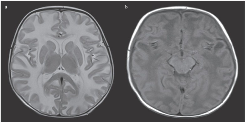
(a) Axial T2 weighted MRI shows bilateral, diffuse, symmetric hyperintense lesions in the cerebral white matter. (b) Axial T1 weighted MRI shows bilateral temporal cysts.

(a) Axial T2 weighted MRI shows bilateral, diffuse, symmetric hyperintense lesions in the cerebral white matter. (b) Axial T1 weighted MRI shows bilateral temporal cysts.