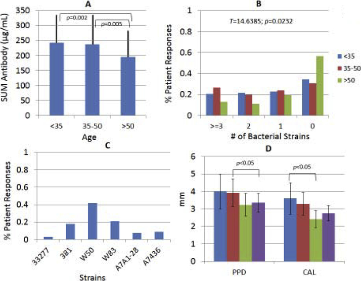
Figure 2A-D:
(A) The overall antibody level to all 6 strains within the different age groups of periodontitis patients. The bars denote the group means and the vertical line is 1 SD. (B) Distribution of antibody reactivities in different age groups in which each patient’s sample was determined to be in the top tertile of antibody of the entire population to each strain. The oldest group had significantly fewer patients with antibody responses in the top tertile across all the P. gingivalis strains. (C) Depiction of the % of patients whose antibody levels were in the top tertile of only 1 strain. (D) Relationship of patients with antibody levels in the top tertile to 0, 1, 2, or ≥3 strains. The bars denote the group means of probing pocket depth and clinical attachment level within the response categories. The vertical brackets denote 1 SD.

