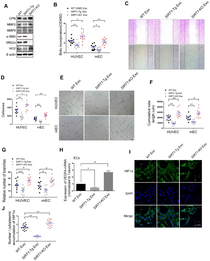
Figure 3.
The exosomes of SIRT1-Tg VSMCs inhibit the endothelial angiogenic functions in vitro. (A) VSMCs from WT, SIRT1-Tg or SIRT1-KO mice were incubated under hypoxia for 24 h. Western blot of OPN, MMP2, MMP9, α-SMA, SM22α and NG2. (B-J) HUVECs or mouse ECs were incubated with the hypoxia-induced VSMC exosomes (Exo) for 24 h and exposed to hypoxia. (B) The relative activity of proliferation by BrdU incorporation. (C and D) The relative activity of migration using a cell-wounding assay. (E) Representative images of tube formation. Scale bars = 200 μm. Relative tube length (F) and number of branches (G) were quantified by measuring the cumulative tube length and branches. (H) qRT-PCR of VEGFA expression in mouse ECs. (I) Immunofluorescent confocal microscopy of HIF1α nuclear translocation in the ECs. Scale bars =100 μm. (J) The quantification of the nuclear-to-cytosol ratio of HIF1α protein in ECs (n=15). Bar graphs show mean±SEM. Student's t-test or one-way ANOVA: *P<0.05, **P<0.01 versus the corresponding control.

