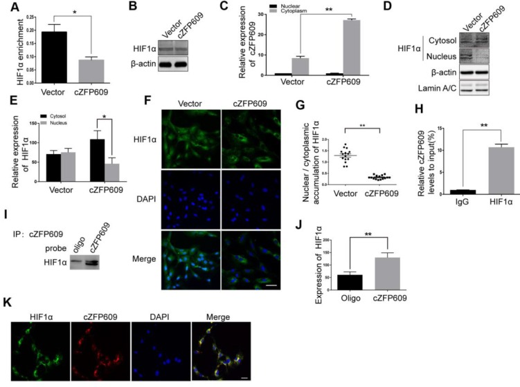
Figure 6.
cZFP609 interacts with and blocks HIF1α nuclear translocation in ECs. The mouse ECs were transfected with vector or cZFP609 for 24 h and then incubated under hypoxia for 24 h. (A) ChIP assay for VEGFA gene promoter region in the ECs using HIF1α antibody. (B) Western blot analysis of HIF1α in the ECs. (C) qRT-PCRs for cZFP609 expression in the nucleus and cytoplasm of the ECs. (D and E) Western blot for HIF1α expression in the ECs. (F) Immunofluorescent confocal microscopy of HIF1α nuclear translocation in the ECs. Scale bars =100 μm. (G) The quantification of the nuclear-to-cytosol ratio of HIF1α protein in ECs (n=15). (H) RIP assay was performed using HIF1α antibodies in the ECs. qRT-PCR was used to detect pulled-down cZFP609. (I and J) The cytoplasm was extracted in ECs incubated under hypoxia for 24 h. RNA pull-down assay was performed using the probe. Western blot was used to validate the interactions between cZFP609 and HIF1α. (K) Confocal FISH images of colocalization between HIF1α and cZFP609 in the ECs. Scale bars=50 μm. Bar graphs show mean±SEM from 3 independent experiments (n=3). Student's t-test: *P<0.05, **P<0.01 versus the corresponding control.

