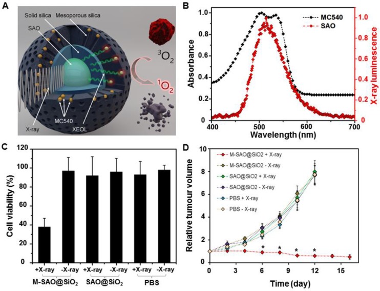
Figure 10.
(A) Schematic illustration of the structure of M-SAO@SiO2 and the working mechanism of SAO-based X-PDT. (B) The XEOL of SAO (red) and the absorption of MC540 (black). (C) Viabilities of U87MG cells with different treatments. (D) Tumor growth curves of mice intratumorally injected with M-SAO@SiO2, SAO@SiO2, and PBS, followed by X-ray irradiation. M-SAO@SiO2: Nanosensitizers consisted of a core made of SrAl2O4:Eu (SAO) and a silica coating loaded with merocyanine 540 (MC540), SAO@SiO2: Silica-coated SrAl2O4:Eu. Adapted with permission from Ref 8. Copyright 2015 American Chemical Society.

