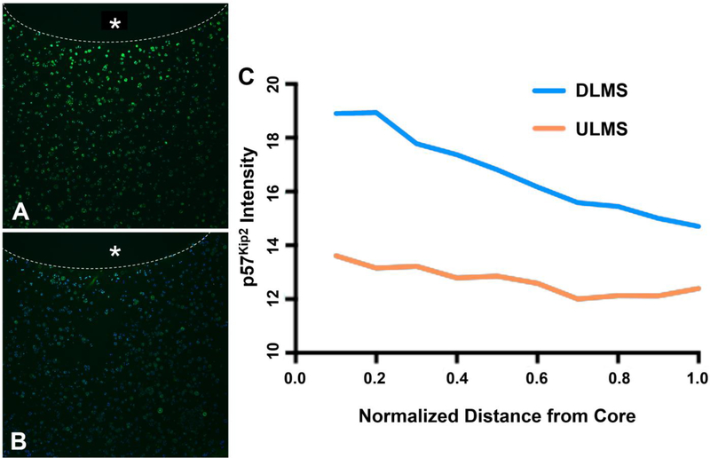
Figure 3.
Representative immunohistochemical stain for p57Kip2 expression (green) counterstained with DAPI. Inner core containing either (A) DLMS or (B) ULMS marked with * and boundary outlined with dotted line; (C) Relative pixel intensity of immunohistochemical stain expression as a function of distance away from the microsphere embedded core (n=1).

