Figure 2:
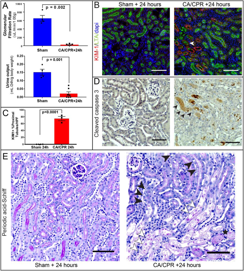
Histopathologic characterization of acute cardiorenal syndrome 24 hours after cardiac arrest and cardiopulmonary resuscitation (CA/CPR). Glomerular filtration (A) and urine output (B) are near zero 24h after CA/CPR. B: Coimmunostaining for Kidney Injury Molecule-1 (KIM-1) and Lotus tetragonolobus lectin (LTA, a marker marker specific for brush borders of proximal tubules) demonstrates heterogeneously distributed injury to proximal tubule cells after CA/CPR, with tubules demonstrating the most KIM-1 expression also containing luminal LTA and brush-border loss (open arrow, example), while other tubular sections demonstrate faint or absent KIM-1 expression. KIM-1 expression, absent from sham, is significant 24h after CA/CPR (C). D: staining for the apoptosis marker cleaved caspase-3 demonstrates caspase-3 positive casts within tubular lumens and scattered tubular cells after CA/CPR and none in sham, suggesting that caspase-mediated apoptotic cell death occurs before 24h after CA/CPR. E: Brush border effacement, tubular cell swelling and vacuolization (closed arrowheads), tubular cell casts (open arrowheads), and tubular proteinaceous casts (star) are present after CA/CPR compared with normal architecture in sham. All scale bars are 100 μm. All column graph bars represent mean±S.E.M.

