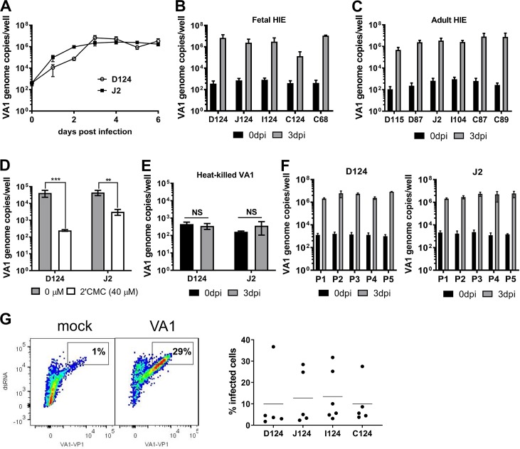
Fig 1. VA1 replicates in human intestinal enteroids (HIE).
A) VA1 growth curve. Undifferentiated D124 and J2 HIE were cultured in 2D monolayers and infected with VA1 (MOI of 1). At indicated times, HIE were harvested and viral RNA was extracted and quantified by RT-qPCR. B-C) Undifferentiated HIE derived from indicated segments of fetal and adult intestines were infected with VA1 and analyzed as before. D) VA1 replication is blocked by the nucleoside analogue 2’-C-methylcytidine (2CMC). Undifferentiated D124 and J2 HIE were infected as before and cells treated with 40 μM 2CMC after adsorption. Cells were harvested and viral RNA quantified at 1 dpi. E) Heat-killed VA1 does not replicate in HIE. VA1 was heat-killed for 3 min at 100°C prior to infection of undifferentiated D124 and J2 HIE. Cells were harvested and viral RNA quantified at 3 dpi. F) VA1 remains infectious for 5 passages in HIE. Supernatant from infected D124 or J2 (passage 1, P1) containing ~103 genome copies/ml was used to infect monolayers of D124 and J2, respectively. Cells were harvested and viral RNA quantified at 3 dpi. The procedure was repeated four times (P2-P5). For all experiments, viral genome copies were measured by RT-qPCR. A–F) Data are from ≥ 3 experiments; error = mean ± SD. G) A single cell suspension of VA1-infected (MOI of 1) D124, I124, J124 and C124 HIE was generated at 3 dpi and stained with antibodies against dsRNA and VA1 capsid protein prior to flow cytometry analysis. The left panel is a representative flow plot of mock- and VA1-infected J124 HIE. The right panel represents the percentage of double-stained cells in each segment. Each dot is a biological replicate. Abbreviations: dpi = days postinfection, D = duodenum, I = ileum, J = jejunum, C = colon. The numbers indicate patient identifiers. **P<0.01; ***P<0.001; NS = not significant.

