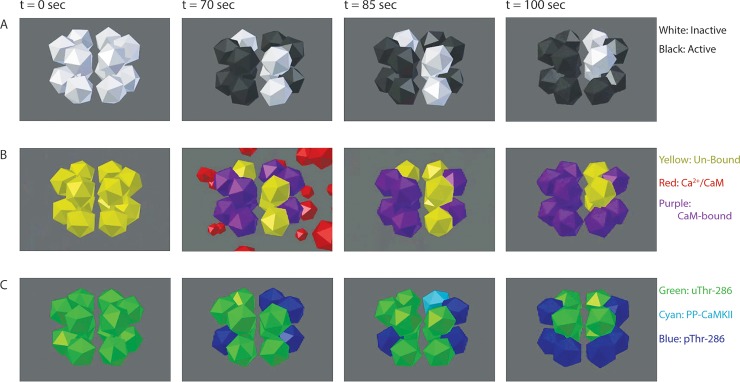
Fig 8. Visualizing individual subunits with MCell and CellBlender.
In the exclusive model, PP does not bind a pThr-286 subunit until Ca2+/CaM dissociation (see t = 85 sec, comparing rows B and C). Each frame depicts the same CaMKII holoenzyme, from the same perspective, at identical time points under 50Hz Ca2+/CaM stimulation. Each dodecahedron is a single CaMKII subunit. (A) Inactive CaMKII subunits (white) spontaneously become active (black) and remain active while bound to Ca2+/CaM. (B) Un-bound CaMKII subunits (yellow) will not bind Ca2+/CaM (red) and become Ca2+/CaM-bound (purple) unless the subunit had previously activated. (C) uThr-286 subunits (green) become pThr-286 (blue). If Ca2+/CaM dissociates from a pThr-286 subunit, then PP can bind and form a PP-CaMKII complex (cyan).

