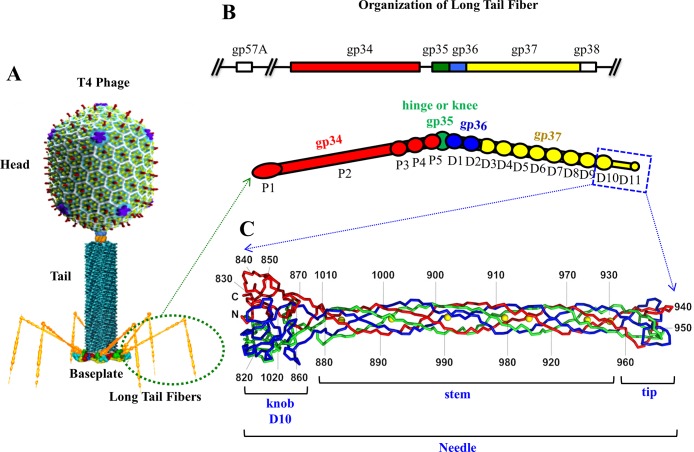
Fig 1. Organization of the bacteriophage T4 long tail fiber.
(A) A structural model of bacteriophage T4 virion showing the head, the tail, and the long tail fibers. (B) The proximal half of the LTF is formed by gp34 trimer (red), the knee cap is formed by gp35 monomer (green), and the distal half is formed by gp36 trimer (blue) and gp37 trimer (yellow) [10]. The part of the gp37 trimer for which the X-ray structure was determined is outlined by blue rectangle. (C) The crystal structure of the gp37 C-terminal fragment [18]. The three polypeptide chains in the gp37 trimer are shown in red, blue, and green. The ferrous ions are shown as yellow spheres.

