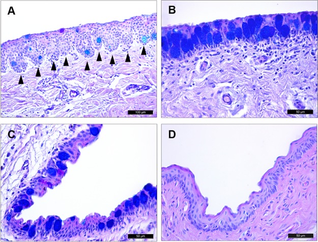
Fig 5. Histological analysis of porcine conjunctiva.
Tissue sections of pig conjunctiva stained with AB/PAS. (A) The marginal conjunctiva between the tarsal and palpebral surfaces was covered by a stratified squamous epithelium and deeper cuboidal epithelial cells. Epithelial downgrowths into the stroma appeared as crypts (arrowheads). (B) The tarsal conjunctiva had a large number of goblet cells containing acidic glycoconjugates. (C) Conjunctiva in the fornix. (D) The bulbar conjunctiva had 4 epithelial cell layers and very few goblet cells.

