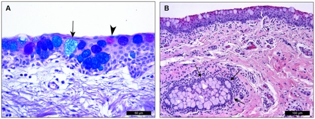
Fig 7. Porcine conjunctival goblet cells.
(A) The different types of goblet cells can be distinguished with AB/PAS staining. Acidic glycoconjugates were stained blue (arrow) by AB, and neutral glycoconjugates were pink (arrowhead) by PAS. Most goblet cells have both types of glycoconjugate granules and appear as dark blue or purple color. (B) H/E staining showed a pseudogland of Henle (arrows) formed by a group of goblet cells embedded within the conjunctival stroma.

