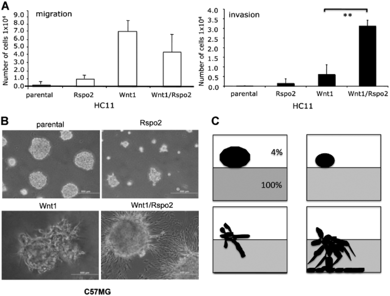
Fig. 7.
Rspo2 and Wnt1 activity in migration and invasion assays. A: 3D Matrigel cultures of parental C57MG cells and cells expressing Rspo2 and Wnt1 alone or together. Wells were covered with 100% Matrigel. Cells were plated in 4% Growth Factors Reduced Matrigel at 3,500 cells/well. Colony formation and Matrigel penetration was determined after 5 days. The original magnification was 100×. The focus was set to detect cells that entered the layer of 100%Matrigel. B: Schematic representation of growth pattern observed in 3D Matrigel culture, corresponding to images in (A). C: Transwell migration and invasion assays of parental HC11 cells and transfectants. HC11 cell pools (2.5 × 10⁵/well) were placed in a transwell migration (upper part) or invasion chamber (lower part), with10%FBS in the lower chamber. The number of cells crossing through the pores in the migration assay was determined at 24 h. The number of cells invading through Matrigel-occluded pores was determined at 48 h. The number of migrating or invading cells is expressed as mean ± SD. **P < 0.02 (t-test). Scale bar = 500 μm.

