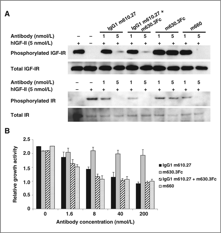
Figure 3.
Inhibition of hIGF-II–mediated IGF-IR and IR phosphorylation (A), and MCF-7 cell growth (B). In the phosphorylation assay, MCF-7 cells were starved in serum-free medium for 5 hours, followed by addition of treatment medium containing 5 nmol/L hIGF-II without or with the antibodies at different concentrations. After 30-minute incubation, cells were chilled and lysed. IGF-IR or IR was immunoprecipitated, and the phosphorylated receptor was detected with a phospho-tyrosine–specific antibody. The membranes were stripped and reprobed by the same polyclonal antibody used for the immunoprecipitation to detect the total amount of the receptors. In the cell growth assay, mean relative light units (RLU) for duplicate wells were determined. Relative growth activity of the cells was calculated by the following formula: average RLU of hIGF-II–containing wells/average RLU of hIGF-II–free wells.

