Figure 3.
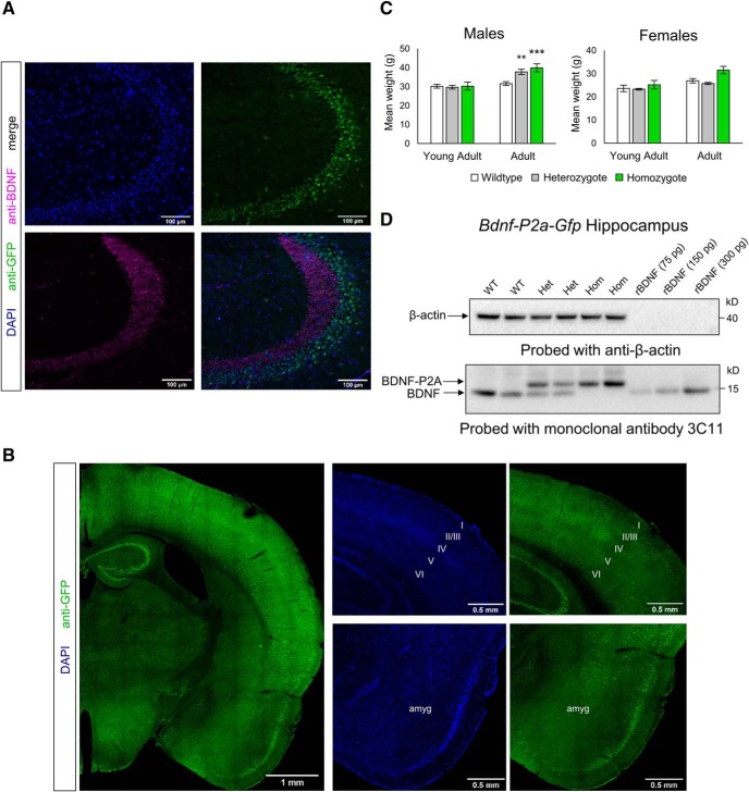
Characterization of Bdnf-P2a-Gfp mice. A, Co-staining of BDNF and GFP homozygous Bdnf-P2a-Gfp hippocampus. Note the clear separation of GFP and BDNF in the mossy fiber projections of hippocampal CA3. B, GFP staining of homozygote brains reveals a comparable staining pattern to previous in situ hybridization experiments, with staining in distinct cortical layers, hippocampal formation, and amygdala. C, Body weights of young adult (3- to 4-month old) and adult (6- to 7-month old) Bdnf-P2a-Gfp mice. While there were no significant differences observed between littermates during young adulthood, significant weight gain could be observed in both heterozygous and homozygous males by 6–7 months of age (p = 0.0100 and p = 0.0017, respectively). The bars represent the mean weights ± SE, n ≥ 7 across genotypes and age categories. D, Western blot analysis of adult Bdnf-P2a-Gfp brain lysates. Note the shift in the molecular weight of BDNF after the addition of the P2A sequence, and the separation of BDNF-P2A from GFP in Bdnf-P2a-Gfp heterozygous (Het) and homozygous (Hom) animals (two animals shown per genotype).

