Figure 5.
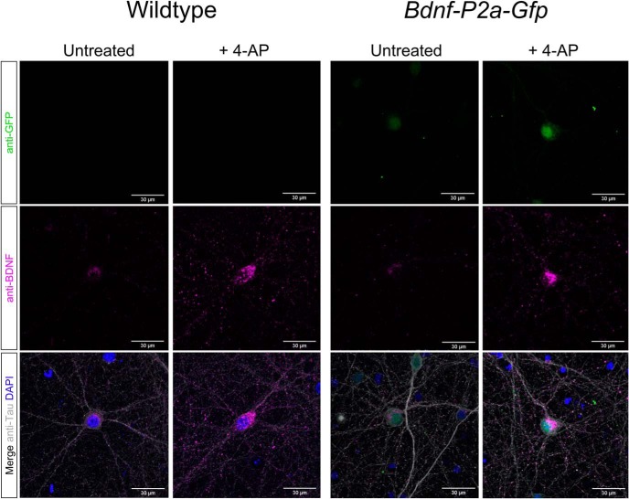
BDNF localization in wild-type versus homozygous Bdnf-P2a-Gfp neurons. Immunostaining of primary neurons with antibodies against BDNF (mAb #9), GFP, and Tau. After 24 h of treatment with 4-AP, note the increased number of BDNF puncta in neuronal projections and the increased GFP signal intensity in Bdnf-P2a-Gfp cultures. Quantification of immunostained Bdnf-P2a-Gfp cultures revealed significant increases in both BDNF and GFP following 4-AP treatment. Quantification of immunostained Bdnf-P2a-Gfp cultures revealed significant increases in both BDNF and GFP following 4-AP treatment (p = 3.94 × 10⁻²¹ and 7.85 × 10⁻²⁴ respectively; n = 90 for both conditions).

