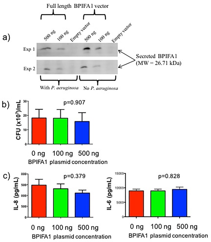
Fig 4. Quantification of colony forming units and inflammatory cytokine production in IB3-1 cells transfected with BPIFA1 plasmid.
IB3-1 cells were transfected with empty vector, 100 ng or 500 ng of BPIFA1 plasmid, and stimulated with P. aeruginosa for 5 hours. This was followed by a) detection of secreted BPIFA1 in transfected IB3-1 cells, b) quantification of colony forming units, and c) quantification of IL-8 and IL-6 production by ELISA. Values represent mean plus standard error of the mean. The Kruskal-Wallis test was used to determine statistical differences between conditions and the corresponding P values are shown.

