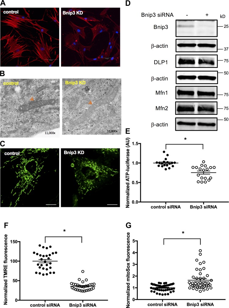
Fig. 4.
F-actin and mitochondrial functions were compromised in Bcl-2 adenovirus E1B 19 kDa-interacting protein 3 (Bnip3) siRNA-transfected human airway smooth muscle (ASM) cells. A: human ASM cells were transfected with Bnip3 and scrambled siRNA for 72 h. Cells were stained with rhodamine-phalloidin and DAPI. Representative images of actin stress fiber were acquired by confocal microscopy at excitation of 540 nm. Bnip3 KD, Bnip3 knockdown. Bar = 100 μm. B: representative transmission electron microscopy images (×11,000) showing mitochondrial structure in control and Bnip3 knockdown ASM cells (red arrow). C: representative confocal live-cell image of mitochondria labeled by MitoTracker green. Bar = 20 μm. D: Western blot for determining protein levels of Bnip3, Mfn1, Mfn2, and DLP1 using β-actin as loading control. E–G: mitochondrial functions were assessed by determining cellular ATP levels (E) using a luciferase assay (n = 18 measurements from 6 different ASM cell lines, *P < 0.05, significance relative to scrambled siRNA), mitochondrial membrane potential (F) using tetramethyl rhodamine ester (TMRE) by confocal live-cell imaging (n = 29 cells from 3 separate experiments; *P < 0.05, significance relative to scrambled siRNA), and mitochondrial reactive oxygen species (G) by confocal live-cell imaging using MitoSox red (n = 43 cells from 3 different experiments; *P < 0.05, significance relative to scrambled siRNA). AU, arbitrary units.

