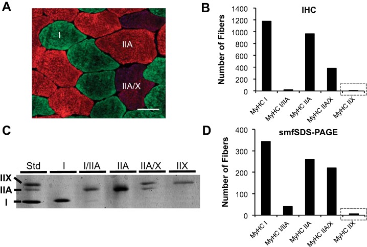
Fig. 1.
Fluorescent immunohistochemistry (IHC; A and B) and single-muscle fiber sodium dodecyl sulfate-polyacrylamide gel electrophoresis (smfSDS-PAGE; C and D) for myosin heavy chain (MyHC) fiber typing in vastus lateralis biopsy samples from a cohort of healthy adult men and women (n = 22; 10 men/12 women). A: representative IHC image of MyHC I (green), MyHC IIA (red), and MyHC IIA/IIX (blue/red) fibers. Scale bar, 50 µm. B: quantification of muscle fiber type via IHC (2,552 total fibers), with gray box emphasizing low MyHC IIX abundance. C: representative image of a smfSDS-PAGE gel showing the continuum of MyHC fiber types. D: quantification of smfSDS-PAGE data from mechanically dissected single muscle fibers (876 total fibers) from the same 22 subjects analyzed in B, with gray box emphasizing low MyHC IIX abundance. Skeletal muscle biopsy samples were obtained under resting conditions, and smfSDS-PAGE was conducted as described by our laboratories in detail elsewhere (36). IHC was conducted in the Toth laboratory according to the methods described in the text, and images were captured at ×20 magnification. Std, standard.

