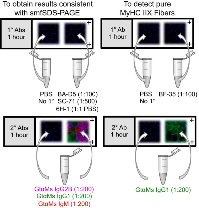
Fig. 3.

Visual representations of recommended immunohistochemistry (IHC) fiber-typing protocols. Muscle fiber borders (laminin, blue) are shown on all images for illustrative purposes. Left: pink is myosin heavy chain (MyHC) I, green is MyHC IIA, and red is MyHC IIX. Right: green is MyHC I and/or IIA, and no fluorescence is MyHC IIX.
