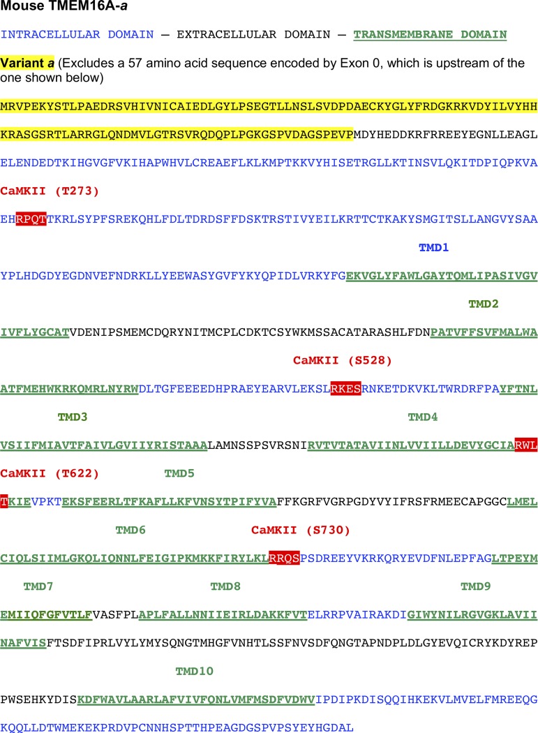
Fig. 1.
Mouse anoctamin-1 sequence used in our study (TMEM16A-a) with the position of the four predicted calmodulin-dependent protein kinase II (CaMKII) sites (RXXT/S). The mouse clone used in our study only included splice variant “a.” The position of the four hypothetical CaMKII phosphorylation sites shown in white font over a red background are relative to the National Center for Biotechnology Information protein database sequence NP_848757.4. Intracellular and extracellular domains are shown in blue and black fonts, respectively. Transmembrane domains are labeled with underlined green fonts. The sequence of splice variant “a” is shown in black font over a yellow background. TMDX, transmembrane domain X. The position of the transmembrane domains is based on the recent Cryo-EM study by Paulino et al. (30) revealing the quaternary structure of mouse TMEM16A.

