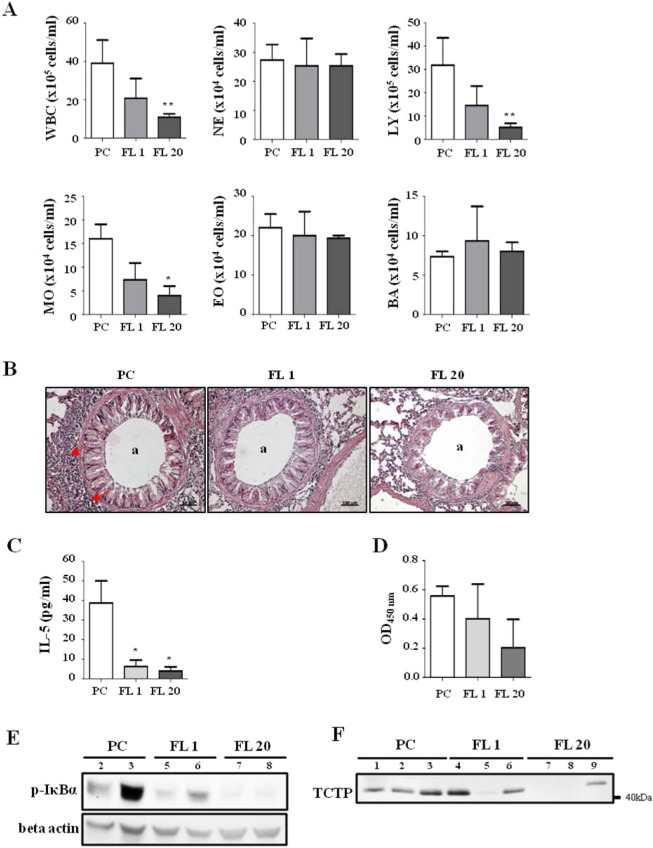
Figure 5.
Dose-dependence of anti-inflammatory effect of FL peptide. (A) Inflammatory cells that infiltrated to the bronchus were measured in BALF using HEMAVET 950FS. (B) H&E staining of lung tissues was performed to visualize cell infiltration. The a indicates the airway, and red arrows indicate inflammatory infiltrates. (C) IL-5 level in BALF was measured using ELISA. (D) OVA-specific IgE in serum was measured using ELISA. (E) Lung tissue was homogenized and immunoblotted with phospho IκBα and beta actin antibodies. (F) BALF was concentrated and immunoblotted for TCTP. Each lane represents biological replicate indicated by the number. PC: positive control (n = 3), FL 1: FL 1 mg/kg (n = 3), FL 20: FL 20 mg/kg (n = 3), WBC: white blood cells, NE: neutrophils, LY: lymphocytes, MO: monocytes, EO: eosinophils, BA: basophils. Values represent mean ± SEM, *p < 0.05, **p < 0.01; compared to PC.

