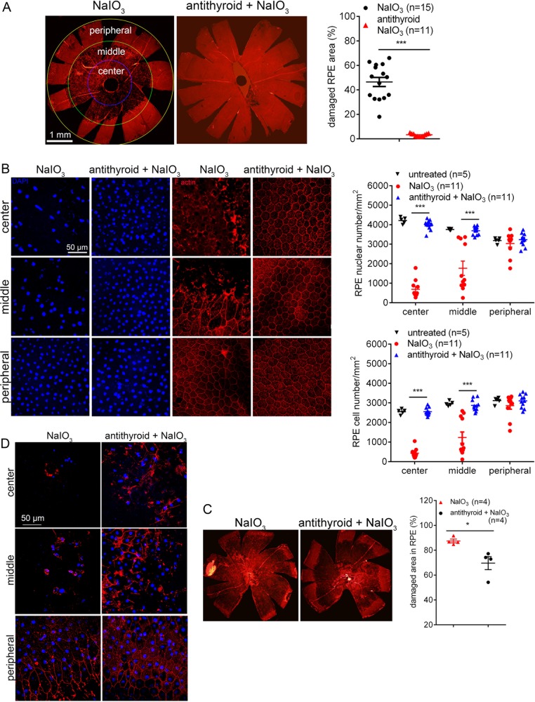
Fig. 1. Treatment with anti-thyroid drug protected RPE from damage and cell loss induced by NaIO3.
RPE morphology and cell loss were evaluated by phalloidin staining for F-actin and DAPI staining for nucleus on RPE whole mounts at 2–3 days post-NaIO3 injection. a, b Shown are RPE morphology evaluations in P30 mice. a Shown are representative low magnification images of phalloidin staining and corresponding quantitative analysis of the damaged area in the RPE. b Shown are representative high magnification images of phalloidin staining and DAPI labeling taken at different regions of the RPE, and corresponding quantitative analysis of RPE cell numbers and RPE nuclear numbers. Data represented the mean ± SEM for 5–15 mice per group (***p < 0.001). c, d Shown are RPE morphology evaluations in 17-month-old mice. c Shown are representative low magnification images of phalloidin staining and corresponding quantitative analysis of the damaged area in the RPE. d Shown are representative high magnification images of phalloidin staining and DAPI labeling taken at different regions of the RPE. Data represented the mean ± SEM for four mice per group (*p < 0.05).

