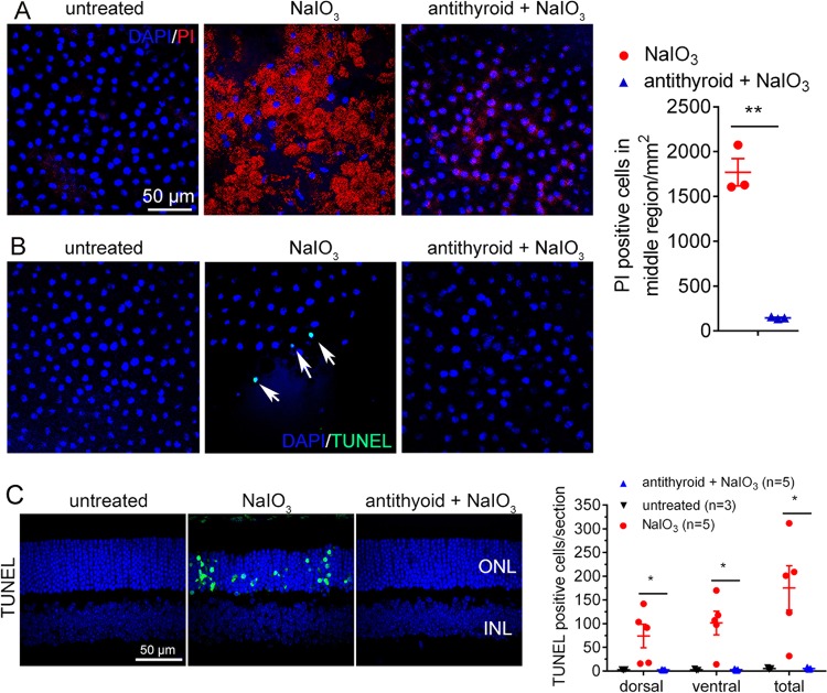
Fig. 3. Treatment with anti-thyroid drug protected RPE cells and photoreceptor cells from necroptosis induced by NaIO3.
a, b RPE cell necroptosis was evaluated by PI staining and TUNEL on the RPE whole mounts. Shown are representative images of PI staining at the middle region of the RPE at 2 days post-NaIO3 injection and correlating quantitative analysis (a), and representative images of TUNEL at the middle region of the RPE at 1 day post-NaIO3 injection (b). c Photoreceptor cell apoptosis was evaluated by TUNEL on the retinal sections at 3 days post-NaIO3 injection. Shown are representative images of TUNEL and correlating quantitative analysis. ONL, outer nuclear layer; INL, inner nuclear layer. Data represented the mean ± SEM for 3–5 mice per group (*p < 0.05, **p < 0.01).

