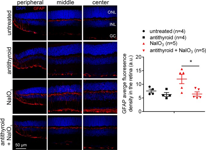
Fig. 5. Treatment with anti-thyroid drug suppressed Müller glia activation induced by NaIO3.
GFAP immunofluorescence labeling was performed on the retinal cross sections at 3 days post-NaIO3 injection. Shown are representative confocal images of immunofluorescence labeling of GFAP on the peripheral, middle, and central regions of the retinal sections and corresponding quantification of immunofluorescence intensity. ONL, outer nuclear layer; INL, inner nuclear layer; GC, retinal ganglion cell. Data represented the mean ± SEM for 4–5 mice per group (*p < 0.05).

