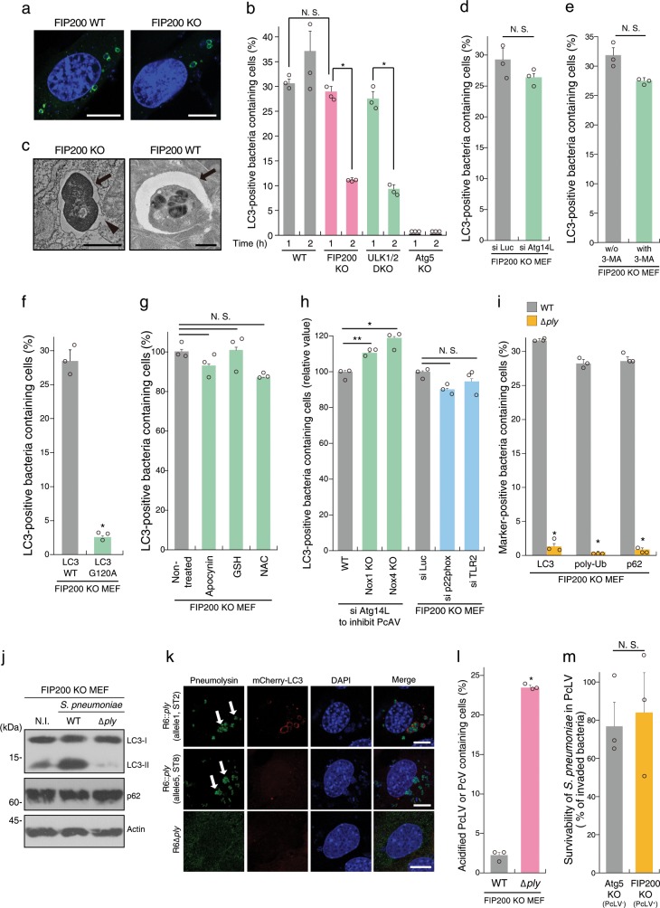
Fig. 1. Streptococcus pneumoniae is engulfed in FIP200-, PI3P-, ROS-independent LAPosome-like vacuoles during early stage of infection.
a Indicated MEFs/GFP-LC3 infected with pneumococci for 1 h were stained with DAPI. b Indicated MEFs/GFP-LC3 infected with pneumococci for 1 or 2 h and stained with DAPI, and percentages of PcLV-containing cells were quantified. c Micrographs of FIP200 KO MEFs at 1 h p.i. or FIP200 WT at 2 h p.i.; Bar, 1 µm. Arrows indicate PcLV or PcAV, and arrowhead indicates ruptured PcLV. d Indicated MEFs/GFP-LC3 treated with indicated siRNAs were infected with pneumococci for 1 h and stained with DAPI, and percentages of PcLV-containing cells were quantified. e FIP200 KO MEFs/GFP-LC3 infected with pneumococci for 1 h with or without 3-methyladenine were stained with DAPI, and percentages of PcLV-containing cells were quantified. f FIP200 KO MEFs/GFP-LC3 G120A infected with pneumococci for 1 h were stained with DAPI, and percentages of PcLV-containing cells were quantified. g FIP200 KO MEFs/GFP-LC3 infected with pneumococci for 1 h with or without indicated antioxidants were stained with DAPI, and percentages of PcLV-containing cells were quantified. h Indicated MEFs/GFP-LC3 treated with indicated siRNA were infected with pneumococci for 1 h and stained with DAPI, and percentages of PcLV-containing cells were quantified. i FIP200 KO MEFs/GFP-LC3 infected with indicated pneumococcal strains for 1 h were stained with DAPI or antibodies against pneumococci, and anti-poly-Ub or -p62 antibodies, and percentages of LC3-, poly-Ub-, or p62-positive bacteria containing cells were quantified. j Lysates from FIP200 KO MEFs infected with indicated pneumococcal strains for 1 h were subjected to immunoblotting with indicated antibodies. k FIP200 KO MEFs/mCherry-LC3 infected with indicated pneumococcal strains for 1 h were stained with DAPI and antipneumolysin antibody. Bar, 10 µm. Arrows indicate pneumolysin around or in the bacterium. l FIP200 KO MEFs infected with indicated pneumococcal strains for 1 h in the presence of 50 nM LysoTracker were stained with DAPI, and percentages of LysoTracker-positive PcV-containing cells were quantified. m Indicated MEFs were infected with pneumococci for 1 h and intracellular survivability of bacteria was determined by colony forming units (cfu); n = 3. Data are expressed as mean ± SEM; *P < 0.01, **P < 0.05, N.S., not significant. Uncropped blots for (j) can be found in Supplementary Fig. 10.

