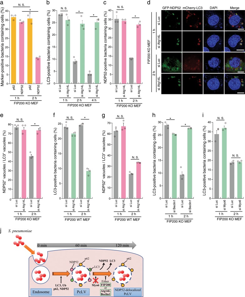
Fig. 3. Atg14L, Beclin1, and FIP200 are involved in NDP52-delocalized PcLVs transition.
a FIP200 KO MEFs/mCherry-LC3 stably expressing GFP-p62 or GFP-NDP52 infected with pneumococci for 1 or 2 h were stained with DAPI, and percentages of each marker-positive bacteria containing cells were quantified. b FIP200 KO MEFs/GFP-LC3 treated with indicated siRNA were infected with pneumococci for 1, 2, or 4 h and stained with DAPI, and percentages of LC3-positive bacteria containing cells were quantified. c–e FIP200 KO MEFs/mCherry-LC3/GFP-NDP52 treated with indicated siRNA were infected with pneumococci for 1 or 2 h and stained with DAPI, and percentages of NDP52-positive bacteria containing cells and the populations of GFP-NDP52+/mCherry-LC3+ vacuoles were quantified. d Representative epifluorescence images in (c). f, g FIP200 WT MEFs/mCherry-LC3 treated with indicated siRNA were infected with pneumococci for 1 or 2 h and stained with DAPI, and percentages of PcLV-containing cells were quantified. h, i FIP200 KO MEFs/GFP-LC3/GFP-NDP52 treated with indicated siRNA were infected with pneumococci for 1 or 2 h and stained with DAPI, and percentages of LC3-positive bacteria containing cells and the populations of GFP-NDP52+/mCherry-LC3+ vacuoles were quantified. j Schematic diagram of PcLV- and subsequent NDP52-delocalized PcLV. Data are expressed as mean ± SEM.; *P < 0.01, N.S., not significant. Bar, 10 µm.

