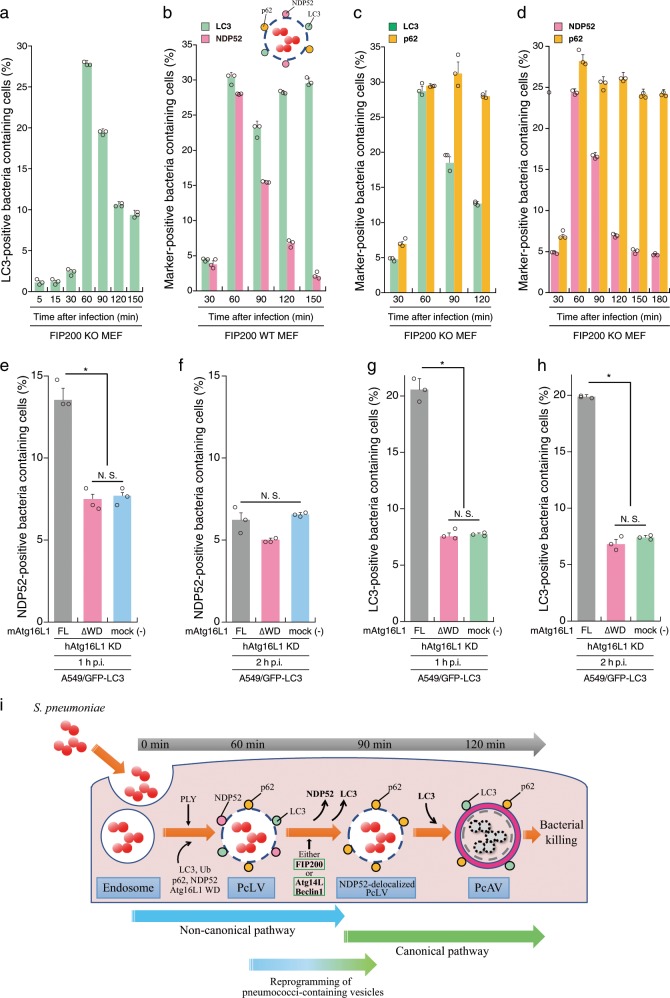
Fig. 4. Hierarchical autophagy processes during time course of pneumococcal infection.
a FIP200 KO MEFs/GFP-LC3 infected with pneumococci for indicated periods were stained with DAPI, and percentages of GFP-LC3 positive bacteria containing cells were quantified. b FIP200 WT MEFs/mCherry-LC3/GFP-NDP52 infected with pneumococci for indicated periods were stained with DAPI, and percentages of each marker-positive bacteria containing cells were quantified. c FIP200 KO MEFs/GFP-LC3 infected with pneumococci for indicated periods were stained with anti-p62 antibody and DAPI, and percentages of each marker-positive bacteria containing cells were quantified. d FIP200 KO MEFs/GFP-NDP52 infected with pneumococci for indicated periods were stained with anti-p62 antibody and DAPI, and percentages of each marker-positive bacteria containing cells were quantified. e–h A549 cells/GFP-LC3/mouse Atg16L1 FL or ∆WD were treated with siRNA for human Atg16L1. After 2 days, the cells were infected with pneumococci for 1 or 2 h, and stained with anti-NDP52 antibody and DAPI, and percentages of each marker-positive bacteria containing cells were quantified. i Schematic diagram of pneumococci-containing endosomes-remodeling to PcAVs through PcLV and NDP52-delocalized PcLV. Data are expressed as mean ± SEM.; *P < 0.01, N.S., not significant.

