Figure 1.
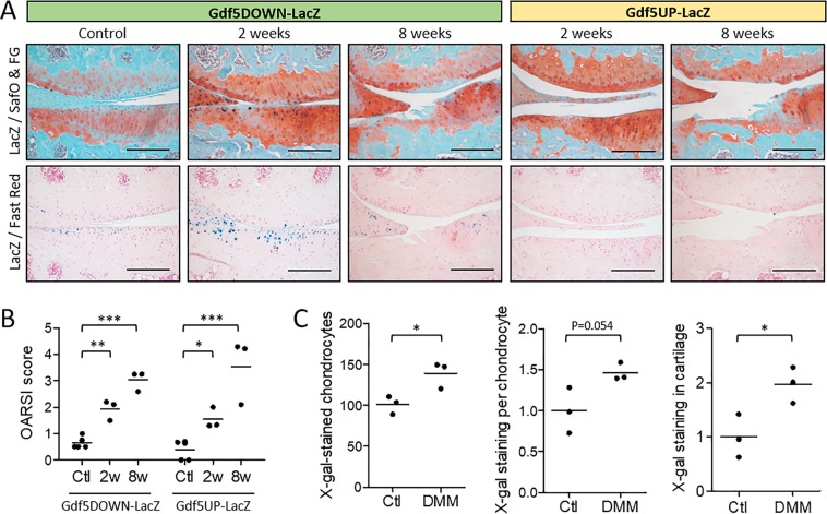
Gdf5 expression in articular cartilage following DMM. (A) LacZ expression (blue X-gal staining) in the articular cartilage of the medial femoral condyle (top of images) and tibial plateau (bottom of images) at 2 and 8 weeks after DMM, or in contralateral control knee, in Gdf5DOWN-LacZ and Gdf5UP-LacZ mice (n = 3 for both strains and both timepoints). At 2 weeks, cartilage shows focal loss of proteoglycan staining (red Safranin O staining) and minor fibrillations at the surface, while at 8 weeks it is severely damaged. LacZ, whole-mount X-gal staining to detect LacZ expression; SafO & FG, Safranin O and Fast Green counterstaining; Fast Red, Nuclear Fast Red counterstaining. Scale bars, 200 μm. (B) OARSI histopathological scores of cartilage damage of the medial tibial plateau at 2 weeks (2w, n = 3) or 8 weeks (8w, n = 3) after DMM surgery, or no surgery (Ctl, n = 5). *p < 0.05; **p < 0.01; ***p < 0.001, two-way ANOVA with Holm-Sidak post-test for comparisons against control. There were no significant differences between the two mouse lines. (C) Number of counted X-gal-stained chondrocytes, average X-gal staining intensity per chondrocyte, and X-gal staining in cartilage calculated by multiplying number and staining intensity of X-gal-stained chondrocytes, in tibial articular cartilage of Gdf5DOWN-LacZ mice at 2 weeks after DMM. Data are expressed relative to the internal contralateral control knees (Ctl). *p < 0.05, two-tailed Student’s t-test.

