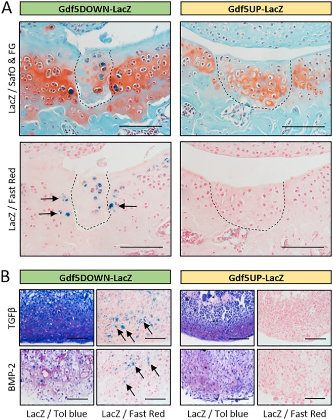
Figure 4.
LacZ expression is upregulated during cartilage repair and in vitro chondrogenesis. (A) Areas of healed cartilage (dashed line) in the patellar groove of the femur of Gdf5DOWN-LacZ (n = 4/10) and Gdf5UP-LacZ (n = 3/10) mice, with LacZ-expressing chondrocytes (blue, arrows) detected in Gdf5DOWN-LacZ mice 4 weeks post-injury. LacZ, whole-mount X-gal staining to detect LacZ expression; SafO & FG, Safranin O and Fast Green counterstaining; Fast Red, Nuclear Fast Red counterstaining. Scale bars, 100 μm. (B) Histological sections of chondrogenic cell pellets. Synovial cells were isolated from Gdf5DOWN-LacZ and Gdf5UP-LacZ mice and treated in vitro for 21 days with TGFβ (10 ng/ml) or BMP-2 (300 ng/ml) to induce chondrogenesis, followed by X-gal staining to detect LacZ expression. Tol blue, Toluidine blue metachromatic staining indicates deposition of cartilage proteoglycans; Fast Red, Nuclear Fast Red counterstaining. LacZ-expressing chondrocytes (blue, arrows) were observed in Gdf5DOWN-LacZ cell pellets, but not Gdf5UP-LacZ pellets, under both culture conditions. Scale bars, 100 μm.

