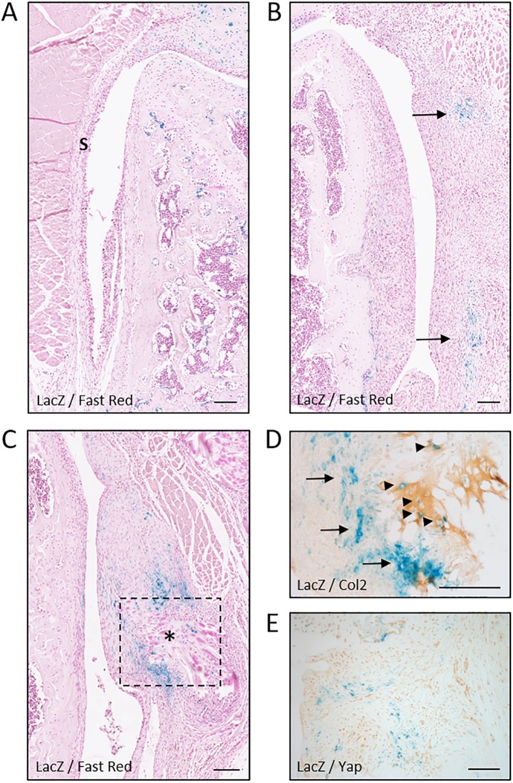
Figure 5.
Gdf5 is switched on in areas undergoing ectopic cartilage formation in synovium. LacZ expression in lateral (A) and medial synovium (B–E) from Gdf5DOWN-LacZ mice 1 week (A,B) or 4 weeks (C–E) after joint surface injury (n = 4 for both timepoints). (A) LacZ expression was not detected in synovium (S) on the lateral side. (B) Clusters of LacZ-expressing fibroblast-like cells (blue, arrows) were found in the medial synovium, near the site of surgical incision. (C) LacZ expression in medial synovium persisted at 4 weeks after injury, particularly near surgical sutures (asterisk). Dotted line indicates area shown in (D) in a consecutive section. (D) IHC staining for Collagen type II (Col2; light brown) revealing LacZ-expressing chondrocytes (blue, arrowheads) embedded in a cartilage matrix surrounded by LacZ-expressing fibroblast-like cells (blue, arrows). (E) IHC staining for Yap showing LacZ-expressing cells (blue) with little or no Yap interspersed between Yap-expressing cells (light brown) that did not detectably express LacZ. LacZ, whole-mount X-gal staining to detect LacZ expression; Fast Red, Nuclear Fast Red counterstaining. Scale bars, 100 μm.

