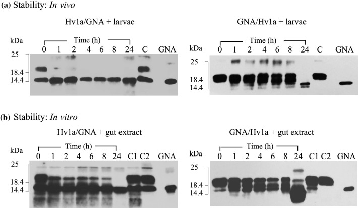
Fig. 4.
Stability of fusion proteins to degradation by A. tumida larval proteases a Western blot analysis of solutions [2.5 mg fusion protein (FP)/ml] in which larvae had been immersed for specified time points. C denotes control FP sample alone (no larvae) incubated at RT for 24 h. b Western blot analysis of FP samples following incubation with larval gut extracts; 75 µg FP + 40 µl gut extract. For each time point, 300 ng FP loaded. C1 denotes control FP alone, C2 is FP + boiled gut extract, both incubated at RT for 24 h. GNA denotes 100 ng recombinant GNA standard

