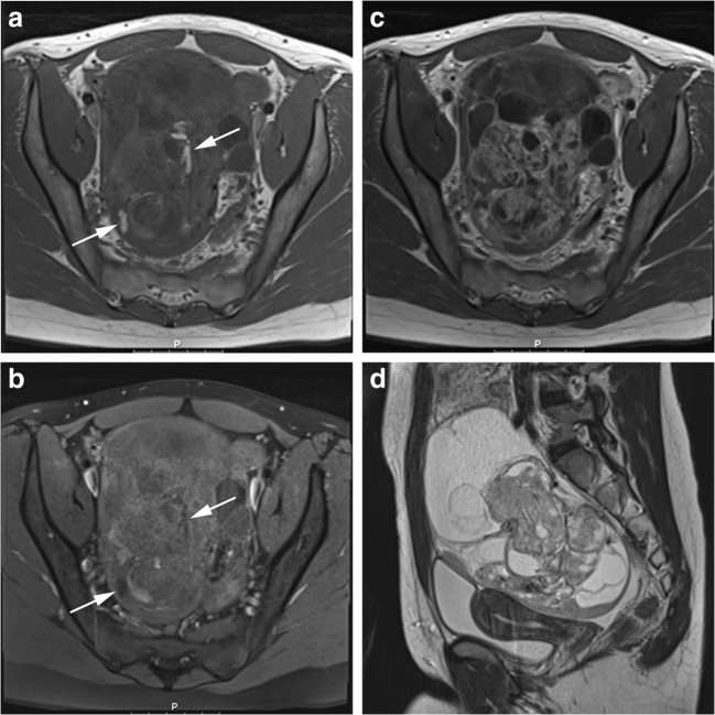
Fig. 1.
An example of immature teratoma grade 1 of the right ovary in a 15-year-old girl, treated as a benign tumour with local resection and follow-up. Axial T1-weighted before and after administration of gadolinium contrast (a, c), axial T1-weighted with fat-suppression (b) and sagittal T2-weighted turbo spin echo (d) show a cystic-solid mass with fatty components (arrows). Intralesional fat is diagnostic for a teratoma. The relative large amount of enhancing parts increases the risk of immature components

