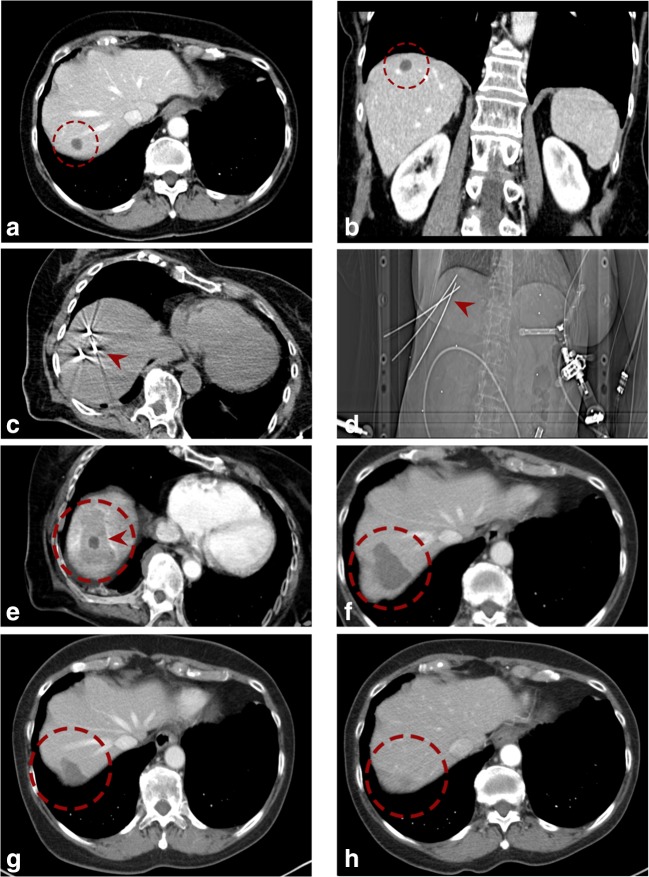
Fig. 2.
Case of a 70-year-old female with a 1.5-cm colorectal cancer liver metastasis in segment VII in the hepatic dome. a, b Portal venous phase initial CT scans with a hypo-enhancing nodule in segment VII (red dashed circle). c, d Axial native control CT and scout (d) with 3 coaxial needles in place (red arrowhead). e Fused CT image of the contrast-enhanced planning and final control CTs showing a complete coverage of the tumor (dark central nodule) by the coagulation zone (red arrowhead). f–h The red dashed circle is marking the progressively shrinking coagulation zone at 3 months (f), 24 months (g), and 48 months (h) after SRFA

