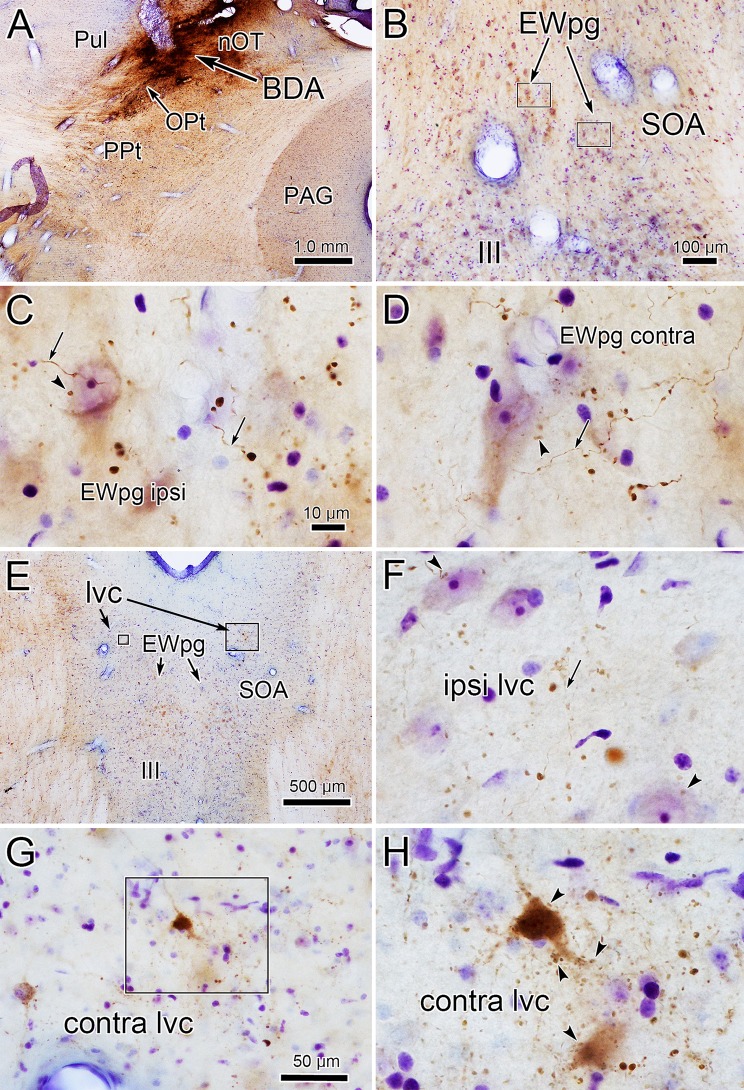
Fig. 7.
Morphology of anterogradely labeled pretectal axons. The BDA injection shown in (a) included the left OPt and spread into the adjacent nOT and Pul. Within the SOA shown in (b), labeled axons (arrows) and boutons were present in both the ipsilateral (ipsi) (c) and contralateral (contra) (d) EWpg, and displayed close associations (arrowheads) with large counterstained neurons. e–h Labeling in the lvc. The area in the small box in (e) is shown in (f) and in the large box is shown in (g). The boxed area in (g) is shown at even higher magnification in (h). Labeled axons (arrow) and terminal fields were present in the lvc ipsilateral (f) and contralateral (g, h) to the injection site. On the ipsilateral side (f), close associations (arrowheads) with counterstained somata are present. On the contralateral side (h), close associations (arrowheads) with retrogradely labeled neurons are present. Scale in c = d, f, h

