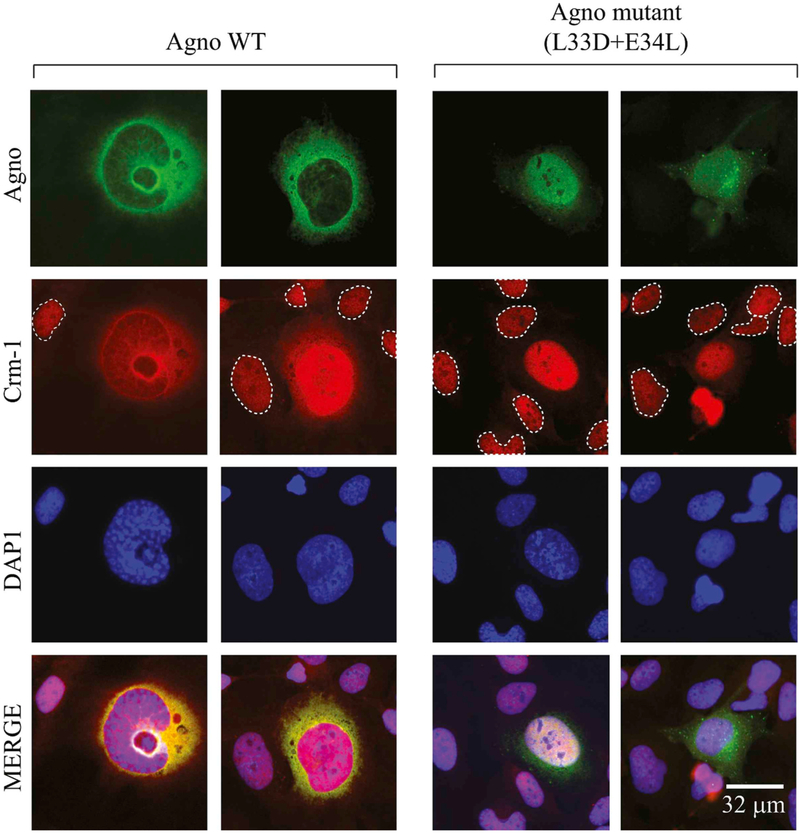
Fig. 8. Analysis of Agno - Crm-1 interactions by immunocytochemistry.
SVG-A cells (Major et al., 1985) were transfected/infected with JCV Mad-1 strain. At 6th day post-infection, the infected cells were fixed with 4% formaldehyde containing 0.05% Triton X-100 and processed for immunocytochemistry in glass slide chambers using primary polyclonal α-Agno and monoclonal α-Crm-1 antibodies and with appropriate secondary antibodies as described in material and methods. The uninfected cells were demarcated by the dashed circles. Cells were finally mounted using mounting medium and examined under a fluorescence microscope (Leica, DMI-6000B, objective: HCX PL APD 60×/1.25 oil, LAS AF operating software) for visualization of the cellular distribution of Agno and Crm-1. Scale bar: 32 μm.

