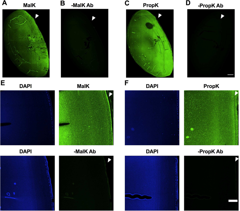
Fig. 1. Immunohistochemical detection of MalK and PropK modified proteins in the human lens.
Immunohistochemical analysis of a human lens transverse sections (donor age: 60 years) showed immunoreactivity (green) for MalK (A) and PropK (C) throughout the lens. Images of higher magnification showed immunoreactivity in both epithelial and cortical fiber cells (E and F top panels). Arrowhead points to the anterior region of the lens. Sections without the primary antibody (negative control) showed no immunoreactivity (B and D as well as bottom panels of E and F). DAPI staining (blue) was used to show the nuclei of epithelial cells. Scale bar = 500 μm for A to D and 100 μm for the images in panels of E and F.

