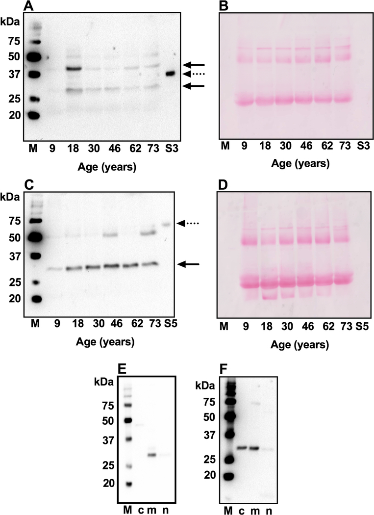
Fig. 5. SIRT3 and SIRT5 in human lens proteins and epithelial cells.
Western blotting of the WS fraction from human lens of varying age using a monoclonal antibody against SIRT3 (A) or SIRT5 (C). The location of SIRT3 and SIRT5 are indicated by arrows. Ponceau staining of western blotted membranes for panels A and C are shown in panels B and D to demonstrate equal protein loading. S3, recombinant SIRT3 and S5, recombinant SIRT5 are indicated by dotted arrows. To identify subcellular localization of SIRT3 and SIRT5 in human lens epithelial cells, the subcellular fractions were western blotted for SIRT3 (E) and SIRT5 (F). Western blots to confirm fractionation into cytosolic, mitochondrial and nuclear fractions are shown in Fig. 4. M, molecular weight markers;

