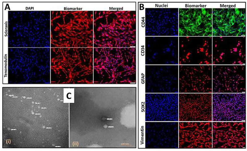
Fig. 1:
(A) Characterization of tenocytes: Immunofluorescence analysis for the expression of Scleraxis and Tenomodulin showing their expression. Images were acquired at 20× magnification using CCD camera attached to the Olympus microscope. (B) Characterization of ADMSCs: Immunofluorescence analysis for the expression of CD44, CD34, GFAP, SOX2 and Vimentin showing their expression. Images were acquired at 20× magnification using CCD camera attached to the Olympus microscope. (C) The examination of the exosome morphology using TEM analysis in tenocytes (i) and ADMSCs (ii) cultured under hypoxic conditions. The tenocytes under hypoxia exhibited more exosomes but smaller size than compared with hypoxic ADMSCs.

