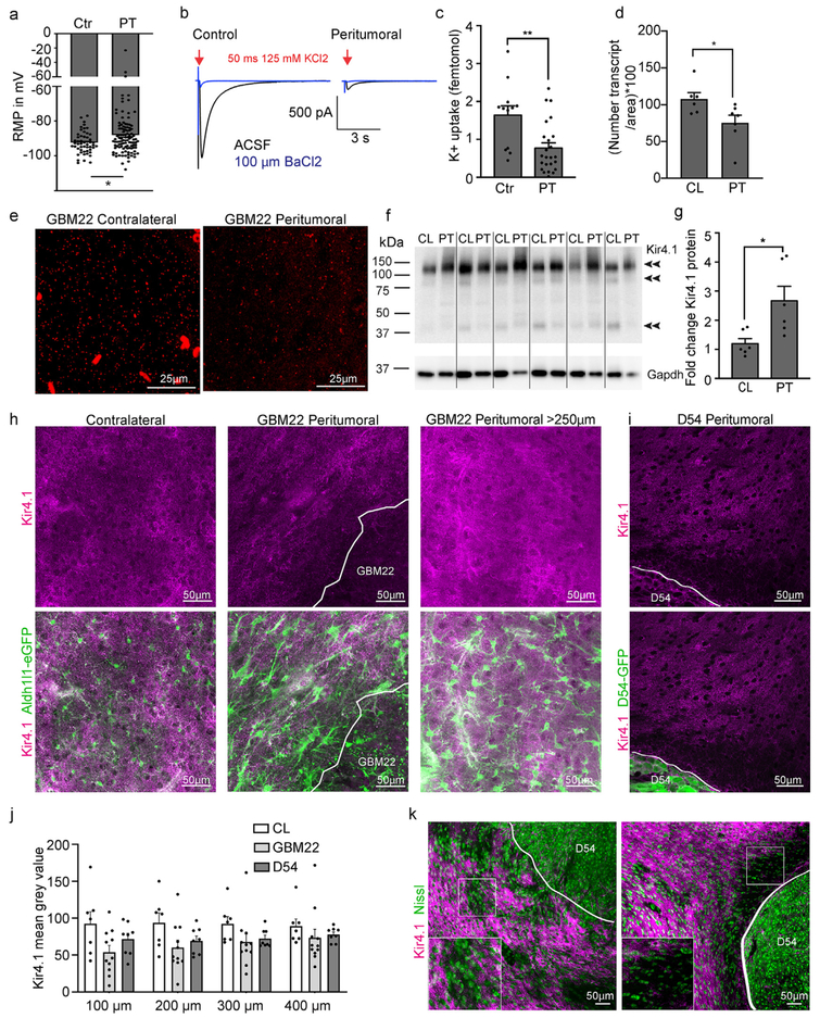
Fig. 5.
Peritumoral astrocytes are impaired in potassium uptake. a. Bar graph showing the resting membrane potential (RMP) of peritumoral and control astrocytes. b. Potassium current traces elicited by a 50 ms puff of 125 mM KCl before (black) and in the presence of 100 μM Ba2+ (blue) in control and peritumoral astrocytes. Cells were voltage-clamped at −80 mV. c. Quantification of potassium uptake. d. Quantification of KNCJ10 (encodes Kir4.1) transcript numbers. e. Representative images of RNAScope (fluorescent in situ hybridization) using probes for KCNJ10. Each small fluorescent dot represents one transcript f,g. Western blot and quantification of Kir4.1 protein in peritumoral cortex compared to the contralateral hemisphere (CL). h. Immunofluorescence and confocal microscopy of Kir4.1 in the peritumoral and contralateral cortex of Aldh1l1-eGFP GBM22 mice. i. Immunofluorescence and confocal microscopy of Kir4.1 in the peritumoral and contralateral cortex of D54 mice. j. Quantification of Kir4.1 signal intensity. k. Double-staining of Kir4.1 and Nissl to assess neuronal density in slices of D54-inoculated mice.

