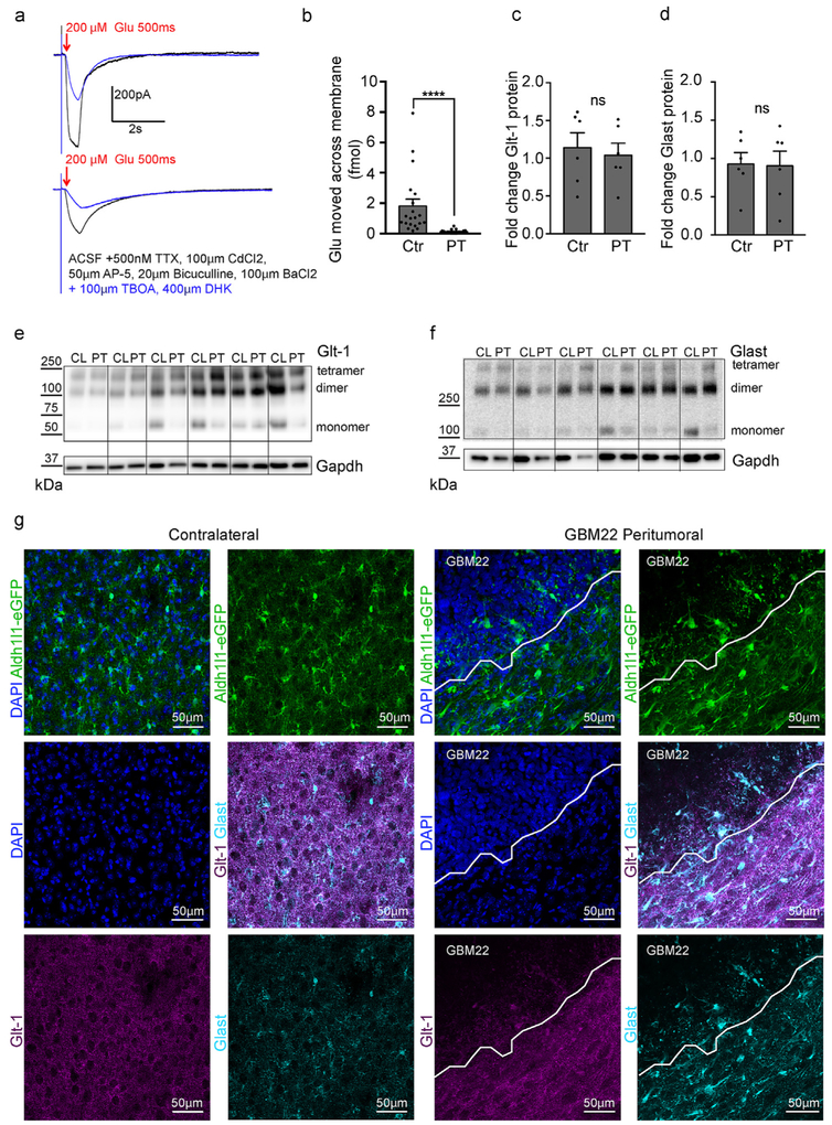
Fig. 6.
Glutamate uptake is impaired in peritumoral astrocytes. a. Recordings of glutamate currents evoked by a 200 μM puff of glutamate for 500 ms from control (top) and peritumoral astrocytes before (black) and after the application of the glutamate transporter inhibitors TBOA and DHK (blue). Cells were voltage-clamped at −80 mV. b. Quantification of the amount of glutamate that moved across the membrane in control and peritumoral astrocytes. c-f. Western blot showing no significant change in Glt-1 (c,e) and Glast (d,f) protein expression in the peritumoral cortex compared to contralateral cortex. g. Confocal imaging of Glt-1 (magenta) and Glast (cyan) expression in contralateral and peritumoral cortex in Aldh1l1-eGFP mice. High density DAPI-positive nuclei demarcate the tumor border (n = 4).

