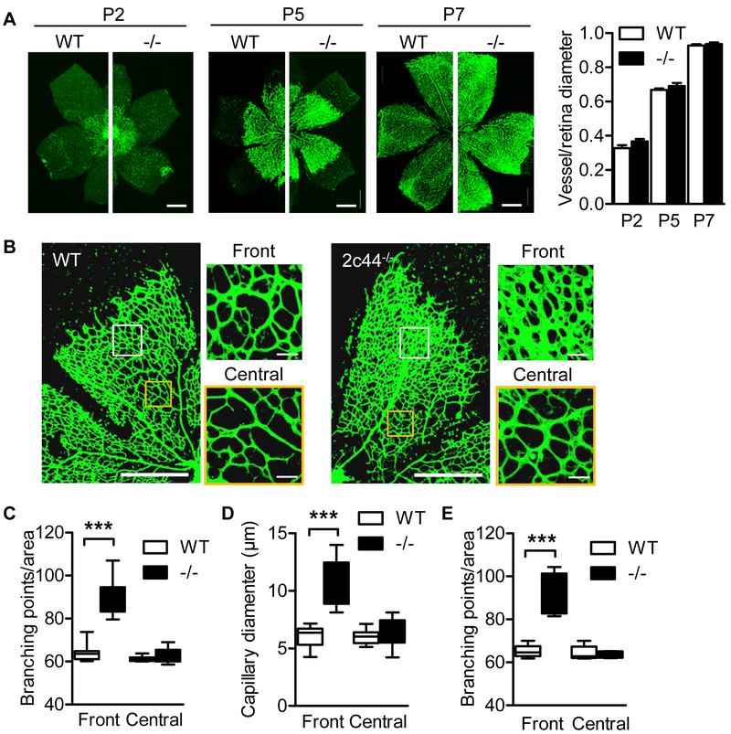
Figure 2. Consequences of Cyp2c44 deletion on retinal angiogenesis.
(A) Isolectin B4 staining was assessed in whole-mounts of the retinal vasculature from wild-type (WT) and Cyp2c44−/− (−/−) mice on postnatal days (P) 2, 5 and 7; bar = 500 μm. (B) Higher magnification images of P5 retinas from wild-type and Cyp2c44−/− mice. The front and central areas indicated by the white and yellow boxes show the position of the right hand images; bars = 500 and 20 μm. (C&D) Quantification of branching points (C) and vessel diameter (D) in P5 retinas from wild-type and Cyp2c44−/− mice. (E) Quantification of branching points in retinas from wild-type and Cyp2c44−/− mice at P7. The graphs summarise data from 6-14 mice in each group; ***P<0.001.

