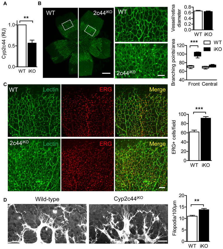
Figure 4. Retinal angiogenesis following the acute knockout of Cyp2c44.
Wild-type (WT) or Cyp2c44iKO (iKO) mice were treated with tamoxifen from P1 to P4. (A) Cyp2c44 expression on P5 determined by RT-qPCR. (B) Isolectin B4 staining in retinal whole mounts from the same animals; bars = 500μm. White boxes indicated the cropped areas shown on the right panels; bar = 100 μm. (C) Quantification of vascularization. (D) Quantification of vessel of branching points in P5 retinas from wild-type and Cyp2c44iKO mice. (E) ERG (endothelial nuclear, red) and Isolectin B4 (green) staining in P5 retinas from wild-type and Cyp2c44iKO mice; bar = 100 μm. (F) High magnification images and quantification of Isolectin B4–stained tip cells and filopodia on P5 retinas. Bar = 20 μm. The graph summarises data from 5-9 mice in each group; **P<0.01, *** P<0.001.

