Figure 5.
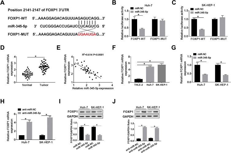
FOXP1 was a target of miR-345-5p. (A) The binding sites of miR-345-5p and FOXP1 3ʹUTR were displayed. (B, C) Dual-luciferase reporter assay was performed to confirm the relationship between miR-345-5p and FOXP1. (D) FOXP1 expression was tested in HCC tissues and adjacent normal tissues. (E) Spearman correlation curve depicted a negative correlation between miR-345-5p and FOXP1 levels in HCC tissues. (F) The level of FOXP1 was measured in THLE-2 cells and HCC cell lines (Huh-7 and SK-HEP-1). (G–J) The mRNA and protein levels of FOXP1 were detected in Huh-7 and SK-HEP-1 cells introduced with miR-NC, miR-345-5p, anti-miR-NC or anti-miR-345-5p by qRT-PCR and Western blot assay. *P < 0.05.

